I do my thing and you do your own. I am not present in this earth to live up to your presumptions, and thus you are not in this world to live up to my own. You're you and I am I, needless to say by chance we discover one another, then its awesome. If not,

Blog

Overview

Acquired adult flatfoot deformity (AAFD) is a progressive flattening of the arch of the foot that occurs as the posterior tibial tendon becomes insufficient. It has many other names such posterior tibial tendon dysfunction, posterior tibial tendon insufficiency and dorsolateral peritalar subluxation. This problem may progress from early stages with pain along the posterior tibial tendon to advanced deformity and arthritis throughout the hindfoot and ankle.Flat Foot


Causes

There are multiple factors contributing to the development of this problem. Damage to the nerves, ligaments, and/or tendons of the foot can cause subluxation (partial dislocation) of the subtalar or talonavicular joints. Bone fracture is a possible cause. The resulting joint deformity from any of these problems can lead to adult-acquired flatfoot deformity. Dysfunction of the posterior tibial tendon has always been linked with adult-acquired flatfoot deformity (AAFD). The loss of active and passive pull of the tendon alters the normal biomechanics of the foot and ankle. The reasons for this can be many and varied as well. Diabetes, high blood pressure, and prolonged use of steroids are some of the more common causes of adult-acquired flatfoot deformity (AAFD) brought on by impairment of the posterior tibialis tendon. Overstretching or rupture of the tendon results in tendon and muscle imbalance in the foot leading to adult-acquired flatfoot deformity (AAFD). Rheumatoid arthritis is one of the more common causes. About half of all adults with this type of arthritis will develop adult flatfoot deformity over time. In such cases, the condition is gradual and progressive. Obesity has been linked with this condition. Loss of blood supply for any reason in the area of the posterior tibialis tendon is another factor. Other possible causes include bone fracture or dislocation, a torn or stretched tendon, or a neurologic condition causing weakness.


Symptoms

Symptoms shift around a bit, depending on what stage of PTTD you?re in. For instance, you?re likely to start off with tendonitis, or inflammation of the posterior tibial tendon. This will make the area around the inside of your ankle and possibly into your arch swollen, reddened, warm to the touch, and painful. Inflammation may actually last throughout the stages of PTTD. The ankle will also begin to roll towards the inside of the foot (pronate), your heel may tilt, and you may experience some pain in your leg (e.g. shin splints). As the condition progresses, the toes and foot begin to turn outward, so that when you look at your foot from the back (or have a friend look for you, because-hey-that can be kind of a difficult

maneuver to pull off) more toes than usual will be visible on the outside (i.e. the side with the pinky toe). At this stage, the foot?s still going to be flexible, although it will likely have flattened somewhat due to the lack of support from the posterior tibial tendon. You may also find it difficult to stand on your toes. Finally, you may reach a stage in which your feet are inflexibly flat. At this point, you may experience pain below your ankle on the outside of your foot, and you might even develop arthritis in the ankle.


Diagnosis

First, both feet should be examined with the patient standing and the entire lower extremity visible. The foot should be inspected from above as well as from behind the patient, as valgus angulation of the hindfoot is best appreciated when the foot is viewed from behind. Johnson described the so-called more-toes sign: with more advanced deformity and abduction of the forefoot, more of the lateral toes become visible when the foot is viewed from behind. The single-limb heel-rise test is an excellent determinant of the function of the posterior tibial tendon. The patient is asked to attempt to rise onto the ball of one foot while the other foot is suspended off the floor. Under normal circumstances, the posterior tibial muscle, which inverts and stabilizes the hindfoot, is activated as the patient begins to rise onto the forefoot. The gastrocnemius-soleus muscle group then elevates the calcaneus, and the heel-rise is accomplished. With dysfunction of the posterior tibial tendon, however, inversion of the heel is weak, and either the heel remains in valgus or the patient is unable to rise onto the forefoot. If the patient can do a single-limb heel-rise, the limb may be stressed further by asking the patient to perform this maneuver repetitively.


Non surgical Treatment

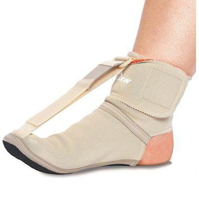
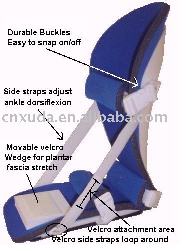
In the early stages, simple pre-fabricated orthotics can help improve the heel position to reduce the mechanical load which is contributing to the symptoms. In advanced stages or long term orthotic use, a plaster of paris or foam box cast can be taken and specific bespoke orthotics manufactured. If the condition develops further a AFO (ankle foot orthotic) may be necessary for greater control. In more advanced stages of symptomatic Adult Acquired flat feet, where the conservative methods of treatment have failed there are various forms of surgery available depending upon the root cause of the issue and severity.

Adult Acquired Flat Foot


Surgical Treatment

If cast immobilization fails, surgery is the next alternative. Treatment goals include eliminating pain, halting deformity progression and improving mobility. Subtalar Arthroereisis, 15 minute outpatient procedure, may correct flexible flatfoot deformity (hyperpronation). The procedure involves placing an implant under the ankle joint (sinus tarsi) to prevent abnormal motion. Very little recovery time is required and it is completely reversible if necessary. Ask your Dallas foot doctor for more information about this exciting treatment possibility.
Overview

Achilles TendinitisThe Achilles tendon attaches the calf muscle to the heel bone. Achilles tendonitis is a repetitive strain (overuse) injury involving lower leg muscles and tendons at the point where they attach to the bone, resulting in pain at the back of the ankle. Chronic overuse can lead to small tears within the tendon causing long-term weakening, making the tendon susceptible to rupture, which could result in a need for surgery.

Causes

Achilles tendinitis is caused by repeated stress to the tendon, not a direct injury. Often times, this can happen from doing too much too soon and not giving your body enough time to rest or adjust to the increase in intensity or amount of exercise. Another contributing factor can be tight calf muscles. Having tight calf muscles and starting an activity can put added stress on the achilles tendon.

Symptoms

The onset of the symptoms of Achilles tendonitis tend to be gradual, with symptoms usually developing over a period of several days, or even weeks. Symptoms may include, Pain, this may be mild at first and may only be noticeable after exercise. Over time the pain may become constant and severe. Stiffness, this is usually relieved by activity. Sluggishness in the leg. Tenderness, particularly in the morning and most commonly felt just above where the tendon attaches to the heel bone. Swelling.

Diagnosis

There is enlargement and warmth of the tendon 1 to 4 inches above its heel insertion. Pain and sometimes a scratching feeling may be created by gently squeezing the tendon between the thumb and forefinger during ankle motion. There may be weakness in push-off strength with walking. Magnetic resonance imaging (MRI) can define the extent of degeneration, the degree to which the tendon sheath is involved and the presence of other problems in this area, but the diagnosis is mostly clinical.

Nonsurgical Treatment

In most cases, nonsurgical treatment options will provide pain relief, although it may take a few months for symptoms to completely subside. Even with early treatment, the pain may last longer than 3 months. If you have had pain for several months before seeking treatment, it may take 6 months before treatment methods take effect. The first step in reducing pain is to decrease or even stop the activities that make the pain worse. If you regularly do high-impact exercises (such as running), switching to low-impact activities will put less stress on the Achilles tendon. Cross-training activities such as biking, elliptical exercise, and swimming are low-impact options to help you stay active. Placing ice on the most painful area of the Achilles tendon is helpful and can be done as needed throughout the day. This can be done for up to 20 minutes and should be stopped earlier if the skin becomes numb. A foam cup filled with water and then frozen creates a simple, reusable ice pack. After the water has frozen in the cup, tear off the rim of the cup. Then rub the ice on the Achilles tendon. With repeated use, a groove that fits the Achilles tendon will appear, creating a "custom-fit" ice pack. Drugs such as ibuprofen and naproxen reduce pain and swelling. They do not, however, reduce the thickening of the degenerated tendon. Using the medication for more than 1 month should be reviewed with your primary care doctor. The following exercise can help to strengthen the calf muscles and reduce stress on the Achilles tendon. Lean forward against a wall with one knee straight and the heel on the ground. Place the other leg in front, with the knee bent. To stretch the calf muscles and the heel cord, push your hips toward the wall in a controlled fashion. Hold the position for 10 seconds and relax. Repeat this exercise 20 times for each foot. A strong pull in the calf should be felt during the stretch. Physical therapy is very helpful in treating Achilles tendinitis. It has proven to work better for noninsertional tendinitis than for insertional tendinitis. Eccentric strengthening is defined as contracting (tightening) a muscle while it is getting longer. Eccentric strengthening exercises can cause damage to the Achilles tendon if they are not done correctly. At first, they should be performed under the supervision of a physical therapist. Once mastered with a therapist, the exercises can then be done at home. These exercises may cause some discomfort, however, it should not be unbearable. Stand at the edge of a stair, or a raised platform that is stable, with just the front half of your foot on the stair. This position will allow your heel to move up and down without hitting the stair. Care must be taken to ensure that you are balanced correctly to prevent falling and injury. Be sure to hold onto a railing to help you balance. Lift your heels off the ground then slowly lower your heels to the lowest point possible. Repeat this step 20 times. This exercise should be done in a slow, controlled fashion. Rapid movement can create the risk of damage to the tendon. As the pain improves, you can increase the difficulty level of the exercise by holding a small weight in each hand. This exercise is performed similarly to the bilateral heel drop, except that all your weight is focused on one leg. This should be done only after the bilateral heel drop has been mastered. Cortisone, a type of steroid, is a powerful anti-inflammatory medication. Cortisone injections into the Achilles tendon are rarely recommended because they can cause the tendon to rupture (tear).

Achilles Tendon

Surgical Treatment

It is important to understand that surgery may not give you 100% functionality of your leg, but you should be able to return to most if not all of your pre-injury activities. These surgical procedures are often performed with very successful results. What truly makes a difference is your commitment to a doctor recommended rehabilitation program after surgery as there is always a possibility of re-injuring your tendon even after a surgical procedure. One complication of surgical repair for Achilles tendon tear is that skin can become thin at site of incision, and may have limited blood flow.

Prevention

So what are some of the things you can do to help prevent Achilles Tendinitis? Warm Up properly: A good warm up is essential in getting the body ready for any activity. A well structured warm up will prepare your heart, lungs, muscles, joints and your mind for strenuous activity. Balancing Exercises, Any activity that challenges your ability to balance, and keep your balance, will help what's called proprioception, your body's ability to know where its limbs are at any given time. Plyometric Training, Plyometric drills include jumping, skipping, bounding, and hopping type activities. These explosive types of exercises help to condition and prepare the muscles, tendons and ligaments in the lower leg and ankle joint. Footwear, Be aware of the importance of good footwear. A good pair of shoes will help to keep your ankles stable, provide adequate cushioning, and support your foot and lower leg during the running or walking motion. Cool Down properly, Just as important as warming up, a proper cool down will not only help speed recovery, but gives your body time to make the transition from exercise to rest. Rest, as most cases of Achilles tendinitis are caused by overuse, rest is probably the single biggest factor in preventing Achilles injury. Avoid over training, get plenty of rest; and prevent Achilles tendinitis.
Plantar Fascia

Overview

Plantar fasciitis was previously believed to be inflammation of the fascia near its insertion on the heel bone. The suffix (-itis) means inflammation. Studies, however, reveal that changes in the tissue associated with the injury are degenerative and not related to inflammation, at least not in the way most people typically think of inflammation. Sudden onset of heel pain may indeed be related to acute inflammation. For persistent heel pain the condition more closely resembles long-standing degeneration of the plantar fascia near its attachment than inflammation. This could explain why anti-inflammatory medications and injections have been unsuccessful at treating it. But there is more to heel pain than just the plantar fascia.


Causes

Factors which may contribute to plantar fasciitis and heel spurs include a sudden increase in daily activities, increase in weight, or a change of shoes or allowing your current shoes to wear excessively. Shoes that are too flexible in the middle of the arch or shoes that bend before the toe joints will cause an increase in tension in the plantar fascia. Make sure your shoes are not excessively worn and that they do not bend in the "middle of the arch".


Symptoms

Patients experience intense sharp pain with the first few steps in the morning or following long periods of having no weight on the foot. The pain can also be aggravated by prolonged standing or sitting. The pain is usually experienced on the plantar surface of the foot at the anterior aspect of the heel where the plantar fascia ligament inserts into the calcaneus. It may radiate proximally in severe cases. Some patients may limp or prefer to walk on their toes. Alternative causes of heel pain include fat pad atrophy, plantar warts and foreign body.


Diagnosis

X-rays are a commonly used diagnostic imaging technique to rule out the possibility of a bone spur as a cause of your heel pain. A bone spur, if it is present in this location, is probably not the cause of your pain, but it is evidence that your plantar fascia has been exerting excessive force on your heel bone. X-ray images can also help determine if you have arthritis or whether other, more rare problems, stress fractures, bone tumors-are contributing to your heel pain.


Non Surgical Treatment

The plantar fascia is a band of tissue, much like a tendon, that starts at your heel and goes along the bottom of your foot. It attaches to each one of the bones that form the ball of your foot. The plantar fascia works like a rubber band between the heel and the ball of your foot to form the arch of your foot. If the band is short, you'll have a high arch, and if it's long, you'll have a low arch, what some people call flatfeet. A pad of fat in your heel covers the plantar fascia to help absorb the shock of walking. Damage to the plantar fascia can be a cause of heel pain.

Plantar Fascitis


Surgical Treatment

Like every surgical procedure, plantar fasciitis surgery carries some risks. Because of these risks your doctor will probably advise you to continue with the conventional treatments at least 6 months before giving you approval for surgery. Some health experts recommend home treatment as long as 12 months. If you can’t work because of your heel pain, can’t perform your everyday activities or your athletic career is in danger, you may consider a plantar fasciitis surgery earlier. But keep in mind that there is no guarantee that the pain will go away completely after surgery. Surgery is effective in many cases, however, 20 to 25 percent of patients continue to experience heel pain after having a plantar fasciitis surgery.


Stretching Exercises

Exercises designed to stretch both your calf muscles and your plantar fascia (the band of tissue that runs under the sole of your foot) should help relieve pain and improve flexibility in the affected foot. A number of stretching exercises are described below. It's usually recommended that you do the exercises on both legs, even if only one of your heels is affected by pain. This will improve your balance and stability, and help relieve heel pain. Towel stretches. Keep a long towel beside your bed. Before you get out of bed in the morning, loop the towel around your foot and use it to pull your toes towards your body, while keeping your knee straight. Repeat three times on each foot. Wall stretches. Place both hands on a wall at shoulder height, with one of your feet in front of the other. The front foot should be about 30cm (12 inches) away from the wall. With your front knee bent and your back leg straight, lean towards the wall until you feel a tightening in the calf muscles of your back leg. Then relax. Repeat this exercise 10 times before switching legs and repeating the cycle. You should practise wall stretches twice a day. Stair stretches. Stand on a step of your stairs facing upstairs, using your banister for support. Your feet should be slightly apart, with your heels hanging off the back of the step. Lower your heels until you feel a tightening in your calves. Hold this position for about 40 seconds, before raising your heels back to the starting position. Repeat this procedure six times, at least twice a day. Chair stretches. Sit on a chair, with your knees bent at right angles. Turn your feet sideways so your heels are touching and your toes are pointing in opposite directions. Lift the toes of the affected foot upwards, while keeping the heel firmly on the floor. You should feel your calf muscles and Achilles tendon (the band of tissue that connects your heel bone to your calf muscle) tighten. Hold this position for several seconds and then relax. Repeat this procedure 10 times, five to six times a day. Dynamic stretches. While seated, roll the arch of your foot (the curved bottom part of the foot between your toes and heel) over a round object, such as a rolling pin, tennis ball or drinks can. Some people find that using a chilled can from their fridge has the added benefit of helping to relieve pain. Move your foot and ankle in all directions over the object for several minutes. Repeat the exercise twice a day.
Plantar Fascia

Overview

Heel pain is most commonly caused by plantar fasciitis, which is the swelling of the tissue that connects the heel bone to the toes at the bottom part of the foot, also known as the plantar fascia. Plantar fasciitis is often described as sharp pain in the middle of the heel, which is typically worse after periods of rest when fascia contracts. The pain usually becomes more bearable as the muscles loosen up during the low-impact walking, but can return even worse after extended periods of walking or standing. The plantar fascia serves as a shock absorber and supports the foot’s arch. Too much tension on the plantar fascia can cause inflammation and swelling. Fortunately in most cases, plantar fasciitis treatment is fairly conservative. Plantar fasciitis exercises, medications and orthotics are usually all that’s needed to manage the pain. Most severe cases may require surgery.


Causes

As a person gets older, the plantar fascia becomes less like a rubber band and more like a rope that doesn't stretch very well. The fat pad on the heel becomes thinner and can't absorb as much of the shock caused by walking. The extra shock damages the plantar fascia and may cause it to swell, tear or bruise. You may notice a bruise or swelling on your heel. Other risk factors for plantar fasciitis include being overweight and obesity. Diabetes. Spending most of the day on your feet. Becoming very active in a short period of time. Being flat-footed or having a high arch.


Symptoms

The most common symptoms of plantar fasciitis include pain on the bottom of the foot near the heel, pain with the first few steps after getting out of bed in the morning, or after a long period of rest, such as after a long car ride. The pain subsides after a few minutes of walking. Greater pain after (not during) exercise or activity.


Diagnosis

After you describe your symptoms and discuss your concerns, your doctor will examine your foot. Your doctor will look for these signs. A high arch, an area of maximum tenderness on the bottom of your foot, just in front of your heel bone. Pain that gets worse when you flex your foot and the doctor pushes on the plantar fascia. The pain improves when you point your toes down. Limited "up" motion of your ankle. Your doctor may order imaging tests to help make sure your heel pain is caused by plantar fasciitis and not another problem. X-rays provide clear images of bones. They are useful in ruling out other causes of heel pain, such as fractures or arthritis. Heel spurs can be seen on an x-ray. Other imaging tests, such as magnetic resonance imaging (MRI) and ultrasound, are not routinely used to diagnose plantar fasciitis. They are rarely ordered. An MRI scan may be used if the heel pain is not relieved by initial treatment methods.


Non Surgical Treatment

Conservative treatment is almost always successful, given enough time. Traditional treatment often includes, rest, NSAIDs, and new shoes or heel inserts. Some doctors also recommend avoiding walking bare-footed. This means you’d have to wear your shoes as soon as you wake up. Certain foot and calf exercises are often prescribed to slowly build strength in the ligaments and muscles that support the arch of the foot. While traditional treatment usually relieves pain, it can last from several months to 2 years before symptoms get better. On average, non-Airrosti patients tend to get better in about 9 months.

Pain In The Heel


Surgical Treatment

Surgery is not a common treatment for this condition. Approximately 5% of people with plantar fasciitis require surgery if non-surgical methods do not help to relieve pain within a year. The surgical procedure involves making an incision in the plantar fascia in order to decrease the tension of the ligament. Potential risks of this surgical procedure include irritation of the nerves around the heel, continued plantar fasciitis, heel or foot pain, infection, flattening of the arch, problems relating to the anesthetic.

Foot Pain

Overview

The plantar fascia is a ligament that connects the heel to the toes on the bottom of the foot. It lies just below the skin layers as it passes over the arch of the foot. A common ailment called plantar fasciitis is the result of this ligament becomes inflamed. This can Foot anatomyhappen from injury, physical stress, or sometimes for no obvious reason. The most common point for this inflammation is where this ligament joints the heel bone. Typical symptoms are the pain on the bottom of the foot near the heel usually most intense in the mornings when arising or after a long period with little movement. The pain typically diminishes with movement. Many suffering from plantar fasciitis have heel spurs. Even though they are in the same area they are unrelated and the heel spurs do not cause the plantar fasciitis. Most times heel spurs will not cause pain and in many go undetected unless they have an x-ray for some other reason.


Causes

You are more likely to develop plantar fasciitis if you are Active, sports that place excessive stress on the heel bone and attached tissue, especially if you have tight calf muscles or a stiff ankle from a previous ankle sprain, which limits ankle movement eg. Running, ballet dancing and aerobics. Overweight. Carrying around extra weight increases the strain and stress on your plantar fascia. Pregnant. The weight gain and swelling associated with pregnancy can cause ligaments to become more relaxed, which can lead to mechanical problems and inflammation. On your feet. Having a job that requires a lot of walking or standing on hard surfaces ie factory workers, teachers and waitresses. Flat Feet or High Foot Arches. Changes in the arch of your foot changes the shock absorption ability and can stretch and strain the plantar fascia, which then has to absorb the additional force. Middle-Aged or Older. With ageing the arch of your foot may begin to sag - putting extra stress on the plantar fascia. Wearing shoes with poor support. Weak Foot Arch Muscles. Muscle fatigue allows your plantar fascia to overstress and cause injury. Arthritis. Some types of arthritis can cause inflammation in the tendons in the bottom of your foot, which may lead to plantar fasciitis. Diabetes. Although doctors don't know why, plantar fasciitis occurs more often in people with diabetes.


Symptoms

Plantar fasciitis is characterized by the following signs and symptoms. Acute plantar fasciitis, pain is usually worse in the morning but may improve when activity continues; if the plantar fasciitis is severe, activity will exacerbate the pain, pain will worsen during the day and may radiate to calf or forefoot, pain may be described anywhere from "minor pulling" sensation, to "burning", or to "knife-like", the plantar fascia may be taut or thickened, passive stretching of the plantar fascia or the patient standing on their toes may exacerbate symptoms, acute tenderness deep in the heel-pad along the insertion of the plantar aponeurosis at the medial calcaneal tuberosity and along the length of the plantar fascia, may have localized swelling. Chronic plantar fasciitis, plantar fasciitis is classified as "chronic" if it has not resolved after six months, pain occurs more distally along the aponeurosis and spreads into the Achilles tendon.


Diagnosis

Most cases of plantar fasciitis are diagnosed by a health care provider who listens carefully to your description of symptoms. During an examination of your feet, your health care provider will have to press on the bottom of your feet, the area most likely to be painful in plantar fasciitis. Because the pain of plantar fasciitis has unique characteristics, pain upon rising, improvement after walking for several minutes, pain produced by pressure applied in a specific location on your foot but not with pressure in other areas, your health care provider will probably feel comfortable making the diagnosis based on your symptoms and a physical examination. Your health care provider may suggest that you have an X-ray of your foot to verify that there is no stress fracture causing your pain.


Non Surgical Treatment

First check your shoes for too much midfoot flexibility and check your training for changes. A detailed evaluation of changes in your training is necessary. You should start with what is called "relative rest" which means a decrease in workout intensity, duration of session and decrease in the number of sessions per week. The most important part of self treatment for this condition is being sure that your shoes offer sufficient stability and are optimal in controlling the forces that contribute to plantar fasciitis and heel spurs. Check your running shoes to make sure that they are not excessively worn. They should bend only at the ball of the foot, where your toes attach to the foot. This is vital! Avoid any shoe that bends in the center of the arch or behind the ball of the foot. It offers insufficient support and will stress your plantar fascia. The human foot was not designed to bend here and neither should a shoe be designed to do this. You should also be doing gentle calf stretching exercises. This will reduce stress on the plantar fascia in two ways. The first manner in which a relaxation of the tension in the calf muscles can help heel pain is that it will reduce the direct pull backwards on the heel bone (calcaneus). The second reason is a little bit more complicated, but essentially it is that a tight achilles tendon and calf muscles causes the rearfoot to move in a manner that causes over pronation as your leg and body move forward over your foot. So go ahead and gently stretch the calf muscle by doing the runner's wall leaning stretch. To strengthen the muscles in your arch toe curls or "doming" can be done. Toe curls may be done by placing a towel on a kitchen floor and then curling your toes to pull the towel towards you. This exercise may also be done without the towel against the resistance of the floor.

Foot Pain


Surgical Treatment

In unusual cases, surgical intervention is necessary for relief of pain. These should only be employed after non-surgical efforts have been used without relief. Generally, such surgical procedures may be completed on an outpatient basis in less than one hour, using local anesthesia or minimal sedation administrated by a trained anesthesiologist. In such cases, the surgeon may remove or release the injured and inflamed fascia, after a small incision is made in the heel. A surgical procedure may also be undertaken to remove bone spurs, sometimes as part of the same surgery addressing the damaged tissue. A cast may be used to immobilize the foot following surgery and crutches provided in order to allow greater mobility while keeping weight off the recovering foot during healing. After removal of the cast, several weeks of physical therapy can be used to speed recovery, reduce swelling and restore flexibility.


Stretching Exercises

The following exercises are commonly prescribed to patients with this condition. You should discuss the suitability of these exercises with your physiotherapist prior to beginning them. Generally, they should be performed 2 - 3 times daily and only provided they do not cause or increase symptoms. Your physiotherapist can advise when it is appropriate to begin the initial exercises and eventually progress to the intermediate and advanced exercises. As a general rule, addition of exercises or progression to more advanced exercises should take place provided there is no increase in symptoms. Calf Stretch with Towel. Begin this stretch in long sitting with your leg to be stretched in front of you. Your knee and back should be straight and a towel or rigid band placed around your foot as demonstrated. Using your foot, ankle and the towel, bring your toes towards your head until you feel a stretch in the back of your calf, Achilles tendon, plantar fascia or leg. Hold for 5 seconds and repeat 10 times at a mild to moderate stretch provided the exercise is pain free. Resistance Band Calf Strengthening. Begin this exercise with a resistance band around your foot as demonstrated and your foot and ankle held up towards your head. Slowly move your foot and ankle down against the resistance band as far as possible and comfortable without pain, tightening your calf muscle. Very slowly return back to the starting position. Repeat 10 - 20 times provided the exercise is pain free.
Foot Pain

Overview

If your first few steps out of bed in the morning cause severe pain in the heel of your foot, you may have plantar fasciitis, an overuse injury that affects the sole of the foot. A diagnosis of plantar fasciitis means you have inflamed the tough, fibrous band of tissue (fascia) connecting your heel bone to the base of your toes.


Causes

When the foot moves, the plantar fascia stretches and contracts. Plantar fasciitis is caused by the repetitive overstretching of the plantar fascia. If the tension on the plantar fascia is too great, this overstretching causes small tears in the plantar fascia. This in turn causes the plantar fascia to become inflamed and painful. Factors that contribute to the development of plantar fasciitis include having very high arches or flat feet, gender, while anyone can develop plantar fasciitis, it tends to occur more commonly in women, exercises such as running, walking and dancing, particularly if the calf muscles are tight. Activities or occupations that involve walking or standing for long periods of time, particularly on hard surfaces, wearing high heeled shoes or shoes that do not offer adequate arch support and cushioning, being overweight, additional weight increases the tension on the plantar fascia, poor biomechanics, extra tension is placed on the plantar fascia if weight is not spread evenly when standing, walking or running. Some cases of plantar fasciitis may be linked to underlying diseases that cause arthritis, such as ankylosing spondylitis.


Symptoms

The typical presentation is sharp pain localized at the anterior aspect of the calcaneus. Plantar fasciitis has a partial association with a heel spur (exostosis); however, many asymptomatic individuals have bony heel spurs, whereas many patients with plantar fasciitis do not have a spur.


Diagnosis

If you see a doctor for heel pain, he or she will first ask questions about where you feel the pain. If plantar fasciitis is suspected, the doctor will ask about what activities you've been doing that might be putting you at risk. The doctor will also examine your foot by pressing on it or asking you to flex it to see if that makes the pain worse. If something else might be causing the pain, like a heel spur or a bone fracture, the doctor may order an X-ray to take a look at the bones of your feet. In rare cases, if heel pain doesn't respond to regular treatments, the doctor also might order an MRI scan of your foot. The good news about plantar fasciitis is that it usually goes away after a few months if you do a few simple things like stretching exercises and cutting back on activities that might have caused the problem. Taking over-the-counter medicines can help with pain. It's rare that people need surgery for plantar fasciitis. Doctors only do surgery as a last resort if nothing else eases the pain.


Non Surgical Treatment

At the first sign of soreness, massage (roll a golf ball under your foot) and apply ice (roll a frozen bottle of water under your foot). What you wear on your feet when you're not running makes a difference. Arch support is key, and walking around barefoot or in flimsy shoes can delay recovery. If pain is present for more than three weeks, see a sports podiatrist. Treatments such as orthotics, foot taping, cortisone injections, night splints, and anti-inflammatories decrease symptoms significantly in about 95 percent of sufferers within six weeks. For more stubborn cases, physical therapy may be prescribed; six months of chronic pain may benefit from shock-wave therapy, an FDA-approved plantar-fasciitis treatment.

Plantar Fascitis


Surgical Treatment

Surgery for plantar fasciitis can be very successful in the right patients. While there are potential complications, about 70-80% of patients will find relief after plantar fascia release surgery. This may not be perfect, but if plantar fasciitis has been slowing you down for a year or more, it may well be worth these potential risks of surgery. New surgical techniques allow surgery to release the plantar fascia to be performed through small incisions using a tiny camera to locate and cut the plantar fascia. This procedure is called an endoscopic plantar fascia release. Some surgeons are concerned that the endoscopic plantar fascia release procedure increases the risk of damage to the small nerves of the foot. While there is no definitive answer that this endoscopic plantar fascia release is better or worse than a traditional plantar fascia release, most surgeons still prefer the traditional approach.


Stretching Exercises

In one exercise, you lean forward against a wall with one knee straight and heel on the ground. Your other knee is bent. Your heel cord and foot arch stretch as you lean. Hold for 10 seconds, relax and straighten up. Repeat 20 times for each sore heel. It is important to keep the knee fully extended on the side being stretched. In another exercise, you lean forward onto a countertop, spreading your feet apart with one foot in front of the other. Flex your knees and squat down, keeping your heels on the ground as long as possible. Your heel cords and foot arches will stretch as the heels come up in the stretch. Hold for 10 seconds, relax and straighten up. Repeat 20 times. About 90 percent of people with plantar fasciitis improve significantly after two months of initial treatment. You may be advised to use shoes with shock-absorbing soles or fitted with an off-the-shelf shoe insert device like a rubber heel pad. Your foot may be taped into a specific position. If your plantar fasciitis continues after a few months of conservative treatment, your doctor may inject your heel with steroidal anti-inflammatory medication. If you still have symptoms, you may need to wear a walking cast for two to three weeks or a positional splint when you sleep. In a few cases, surgery is needed for chronically contracted tissue.
Metatarsal pain, often referred to as metatarsalgia, can be caused by several foot conditions, including Freiberg's disease, Morton's neuroma and sesamoiditis. According to a 2003 article in the British Journal of Sports Flat Feet Medicine,” a flat or high arch is one of many risk factors for lower extremity injuries including foot injuries. Poor circulation occurs when there is not enough blood supplied to an area to meet the needs of the cells.

TOE CONDITIONS: Ingrown toenails, blood accumulation under the nail plate (subungual hematoma), corns and calluses are all often seen as a result of playing baseball. It is important that good foot hygiene be practiced with washing between the toes and drying the feet well after bathing. Topical antifungals work well to treat athletes foot. ORTHOPEDIC INJURIES: Most orthopedic baseball foot and ankle injuries are acute or sudden. If an individuals foot or ankle is injured, seek immediate evaluation with one of our doctors. If your athlete has a baseball related injury, call our specialists at Advanced Foot and Ankle Center in McKinney and Prosper Texas at 972-542-2155. However, toe numbness and pain occurring together is one such problem that you cannot afford to ignore. Common symptoms are flat feet knee problems , burning sensation, numbness.

If you see just a thin line connecting the ball of your foot to your heel, you have high arches. If you have flat feet or high arches, you're more likely to get plantar fasciitis, an inflammation of the tissue along the bottom of your foot. Without proper arch support, you can have pain in your heels, arch, and leg. You can also develop bunions and hammertoes, which can become painful,” says Marlene Reid, a podiatrist, or foot and ankle doctor, in Naperville, IL. Shoes with good arch support and a slightly raised heel can help ward off trouble. Laces, buckles, or straps are best for high arches. See a foot doctor to get fitted with custom inserts for your shoes. Good running shoes, for example, can prevent heel pain, stress fractures , and other foot problems that can be brought on by running. A 2-inch heel is less damaging than a 4-inch heel. If you have flat feet, opt for chunky heels instead of skinny ones, Reid says.Plantar Fasciitis,Pes Planus,Mallet Toe,High Arched Feet,Heel Spur,Heel Pain,Hammer Toe,Hallux Valgus,Foot Pain,Foot Hard Skin,Foot Conditions,Foot Callous,Flat Feet,Fallen Arches,Diabetic Foot,Contracted Toe,Claw Toe,Bunions Hard Skin,Bunions Callous,Bunion Pain,Ball Of Foot Pain,Back Pain

Do not consume food items which you are allergic to. Keep dead skin off your lips by lightly scrubbing them at least twice a week using a mild, natural ingredient such as cornflour or a lemon juice-sugar pack. I had a long road workout two weeks ago and immediately after starting having pain on the ball of my foot in this area. I have also learned buying shoes online is easy.Plantar Fasciitis,Pes Planus,Mallet Toe,High Arched Feet,Heel Spur,Heel Pain,Hammer Toe,Hallux Valgus,Foot Pain,Foot Hard Skin,Foot Conditions,Foot Callous,Flat Feet,Fallen Arches,Diabetic Foot,Contracted Toe,Claw Toe,Bunions Hard Skin,Bunions Callous,Bunion Pain,Ball Of Foot Pain,Back Pain

Went to Podiatrist after receiving pain pills to move, got MRI and he told me I have severe tear in plantor faciitis tendon. Have swelling or what I call a fatty feeling, as I have always had on ball of foot below left most two toes. And it seems to feel a little more fatty since I walked for the first time today after putting on a good pair of ankle boots. Any idea what the fatty feeling is on ball of foot. Lastly, I took the boot off at my stairs into my house 2 days ago and took a step using ball of left foot and it did not pop.
Zetaclear Nail Fungus Treatment is the item for managing nail fungus. Is your cat clawing the furnishings and climbing the curtains? Do you hear a click-click-clicking when your pet is trotting down the hallway toward you? Ranked fifteenth safest in the USA, you'll be guaranteed to want to settle down with this express especially if you have got young children. Dogs may be pets, but for some people dogs are family too.

Lack of sensation in the feet increases the risk for unnoticed foot injuries, which may lead to serious infections. Diabetic peripheral neuropathy can affect the muscle-related, or motor, nerves of the feet and lower legs. Damage to these nerves commonly causes weakness of the associated muscles, which can lead to foot deformities, notes the American College of Foot and Ankle Surgeons on the patient information website Foot Health Facts. Claw toe and hammertoe deformities, in which the toes curl rather than lying flat, frequently occur in people with diabetic motor neuropathy of the feet. These deformities often cause bunions, calluses and corns, which may lead to foot ulcers. The University of Chicago Center for Peripheral Neuropathy reports that pain, tingling or numbness of the hands can occur with diabetic peripheral neuropathy, affecting the sensory nerves of the hands. Claw toes result from an inherent muscle imbalance. A terrific skin care regime.

It basically involves lengthening the tightened tendons responsible for the curled toe Uncurling the toes allows them to rest flat In more severe cases, bits of bone may also need to be chipped away in order to see the toe uncurled. The alignment putter is an improvement on the mallet putter and offers different alignment features that help putt the ball on the target line. The term hammer toe deformity (HTD) refers to the 'buckling' effect of the toes at the joint furthest from the tip of the toe (the proximal interphalangeal joint-between the proximal and intermediate phalanges). Keep your feet busy—use those joints to keep them flexible.

Foot care is an essential routine activity that should be done by all individuals to achieve optimum wellness. Foot is a significant part of your physique therefore, you should value it. You must be mindful that there are lots of approaches to protect yourself from foot ailments. Reflexology is an ancient art of applying pressure and massage to reflex points on the foot. Reflexology treatment is aimed to give complete relaxation and overall benefits through foot reflex manipulation and general foot and lower leg massage. Hammer toe is more likely to simultaneously occur with bunions.Plantar Fasciitis,Pes Planus,Mallet Toe,High Arched Feet,Heel Spur,Heel Pain,Hammer Toe,Hallux Valgus,Foot Pain,Foot Hard Skin,Foot Conditions,Foot Callous,Flat Feet,Fallen Arches,Diabetic Foot,Contracted Toe,Claw Toe,Bunions Hard Skin,Bunions Callous,Bunion Pain,Ball Of Foot Pain,Back Pain

Most patients respond to non surgical treatment such as the prescription of orthoses, but on occasion surgery is required. Heel bumps (Haglund's Deformity) An enlargement of the bone at the back of the heel which can encourage bursitis to develop. Bone spurs An excessive growth of bone causing pain or limitation of movement. Achilles tendon problems Most patients respond to non-surgical treatment. Tendon lengthening is sometimes required to treat the condition. Pain in the region of the heel and a formation of a bump just above the heel indicates Haglund's Deformity. One reason for the affliction is the pressure from the back portion of the shoe on the rear of the heel. Pressure on the heel gets considerably reduced. In an extreme case of surgery may have to done for removal of the bump. A small incision is made on the rear of the heel. A little part of the heel bone is removed. This will ensure Haglund's Deformity that no extra pressure comes on to the main heel bone. Another form of deformity is Bunions. Claw toes.Plantar Fasciitis,Pes Planus,Mallet Toe,High Arched Feet,Heel Spur,Heel Pain,Hammer Toe,Hallux Valgus,Foot Pain,Foot Hard Skin,Foot Conditions,Foot Callous,Flat Feet,Fallen Arches,Diabetic Foot,Contracted Toe,Claw Toe,Bunions Hard Skin,Bunions Callous,Bunion Pain,Ball Of Foot Pain,Back Pain

Marx RM. Anatomy and pathophysiology of lesser toe deformities. Taylor RG. The treatment of claw toes by multiple transfers of flexor into extensor tendons. Topics are richly illustrated with more than 40,000 clinical photos, videos, diagrams, and radiographic images. More than 1000 clinical procedure articles provide clear, step-by-step instructions and include instructional videos and images to allow clinicians to master the newest techniques or to improve their skills in procedures they have performed previously. Typically, the DIP joint is extended in a hammer toe. Claw toe makes your toes bend and look like a claw.

It can also be caused by muscle, nerve, or joint damage resulting from conditions such as osteoarthritis , rheumatoid arthritis , stroke , Charcot-Marie-Tooth disease , Complex Regional Pain Syndrome or diabetes 3 Hammer toe can also be found in Friedreich's ataxia (GAA trinucleotide repeat). The Mayo Clinic refers to it as claw-like” while the American Podiatric Medical Association says that it looks like an inverted V from the side. The skin on top of the joint often becomes irritated from rubbing against the shoe. Sometimes the ball of the foot aches where it meets the affected toe.

First blog

03/14/2014 14:47

Our new blog has been launched today. Stay focused on it and we will try to keep you informed. You can read new posts on this blog via the RSS feed.